


Arthrosis means a pathological process characterized by dystrophy and degeneration of articular cartilage. As a rule, the matter is not limited to cartilage only - then the pathology spreads to the bone tissue (subchondral) located below the cartilage. Therefore, arthrosis is also called osteoarthritis. And because all of these disorders eventually lead to changes in joint structure, this process is called deforming osteoarthritis, which can affect any joint. In clinical practice, in most cases arthrosis of the knee joint or gonarthrosis is observed.
The essence of pathology
In terms of frequency and prevalence, knee arthrosis is second only to hip arthrosis (coxarthrosis). To find out what causes this, it is desirable to consider briefly the anatomical structural features of the knee and the functions it performs. This is one of the largest joints, in the formation of 3 bones involved - the femur, tibia and patella. Thus, it is a complex joint formed by 2 joints - the patellofemoral and patellofemoral joints.
The articular surfaces of all 3 bones are covered with cartilage, which facilitates movement in the joints and protects the subchondral bone tissue from mechanical wear. In addition to the articular cartilage itself, the knee has a paired menisci cartilage formation that enhances the congruence (anatomical correspondence) of the articular surface. Articular cartilage does not have its own blood vessels. Its feeding is carried out diffusely from the intra-articular (synovial fluid). Like a sponge, cartilage shrinks under mechanical stress during movement, carrying heavy loads. During this time, waste products are released from the cartilaginous tissue into the synovial fluid around it. Conversely, at the time of relaxation, rest, synovial fluid and nutrients contained in it penetrate into the cartilage of the knee.
For some reason, the nutrition of the articular cartilage of the knee joint is disrupted, which involves knee arthrosis. At the same time, initially, there is a lack of nutrients in cartilage tissue - chondroitin sulfate, glucosamine, calcium and other micro -elements. Moisture is gone. This is a process of dystrophy, followed by degeneration - thinning of the articular cartilage. In turn, this negative process leads to structural and motor disorders in the knee joint.
Knee arthrosis is often mistaken for salt deposition. Suppose, some mineral salts, including table salt, are stored in microcrystalline form in the articular cavity, leading to pain and movement disorders. This is not true. Apparently, a completely different process is taken for salt deposition. In response to the destruction of articular cartilage in the subchondral bone, marginal bone growth - osteophytes - are formed to stabilize the knee at least to some degree. However, in the future, osteophytes only exacerbate arthrosis, and contribute to further destruction of cartilage.
Cause
The causes of knee joint arthrosis are various, and may be due to the pathology of the knee itself, or other diseases and metabolic disorders. In this case, gonarthrosis can be primary and secondary. The mechanism of primary arthrosis is not fully understood. It is believed that in this case, the disease is caused by a combination of factors, including:
- Old age, when degenerative changes occur not only in articular cartilage, but also in all organs and tissues;
- Excess weight, which increases the mechanical stress on the joints;
- Physical inactivity, or conversely, excessive physical activity;
- Some congenital anatomical disorders of the knee, in which the articular cartilage and subchondral bone are initially altered;
- General metabolic disorders leading to changes in the mineral composition of synovial fluid.
Secondary arthrosis of the knee joint is a complication of another disease. Often, the disease is arthritis of various natures - gouty, rheumatic, rheumatoid, septic, tuberculosis, etc. In this disease, various pathological factors (infection, aberrant immune response, uric acid crystals) form an inflammation of the synovial membrane in the so -called form. synovitis. Synovitis is necessarily accompanied by a deterioration in the quality of synovial fluid, which in turn, leads to arthrosis.
Another common cause of arthrosis is a knee injury. Post-traumatic arthrosis of the knee joint is the result of intra-articular fractures of the femur and tibia, hemarthrosis (joint bleeding), damage to the knee ligaments and menisci. Here, the pathology is based on mechanical factors (damage) and the damage that develops subsequently (arthritis). In addition, osteoporosis is often accompanied by arthrosis. Lack of calcium in bones requires the destruction of not only bone, but also cartilage tissue.
symptoms
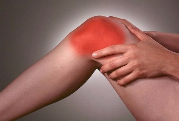
The main symptoms of knee joint arthrosis:
- pain;
- Impaired knee movement;
- Difficulty walking;
- Crusting when moving;
- First - pathological tension, and then - muscle atrophy of the lower limbs;
- Defects of the knee joint.
Initially, as a rule, the patellofemoral joint suffers, which accounts for most of the functional load. In general, knee pads with arthrosis are probably the most vulnerable. It is from the cartilage of the kneecap that dystrophic changes begin in arthrosis. Clinically, this is indicated by swelling and pain when feeling these bones. As a result of dystrophic changes, the articular cartilage undergoes sclerotic changes - it loses its elasticity, is replaced by coarse connective tissue.
Subsequently, the articular bag and ligament apparatus undergo sclerotic changes. Joint configuration changed. Initially, due to the corresponding arthritis, it is swollen, inflamed. Subsequently, with the development of degeneration and sclerosis, the amount of synovial fluid decreases dramatically, the joint space narrows, which inevitably leads to movement disorders. At first, the gait is difficult and the limb muscles become tense. Then ankylosis develops - complete immobility of the knee, and, consequently, atrophy of the muscles of the thighs and lower legs. All of these changes take shape over a long period of time. In this case, there are 3 degrees of arthrosis:
- Arthrosis of the knee joint degree 1. The pain is localized mainly in the area of the kneecap and along the inner surface of the knee joint. Pain is of a "beginning" nature - it appears at the beginning of the movement, and then subsides. Also, pain can appear with significant effort (walking long distances, carrying loads), and disappear after rest. There is no structural change in the joint at this stage.
- Stage 2 knee joint arthrosis. Pain can occur even during rest, and bother for longer periods of time. Limits of range of motion (contractures) appear in the knee. The patient was lame, forced to move with a stick. Inflammatory and dystrophic changes in the joints are formed, externally indicated by an increase in the knee due to edema.
- Stage 3 knee joint arthrosis. Severe knee pain that does not stop even after a long rest. Severe irreversible disruption in the joint structure, leading to ankylosis and loss of ability to move. Changes in the configuration of the entire lower limb, indicated by the curvature of the valgus or varus (O or X -shaped).
The diagnosis of knee arthrosis is carried out on the basis of the above symptoms and complaints of patients, as well as X-ray data (narrowing of the joint space, osteophytes, osteoporosis, hardening of the bones). Gonarthrosis is treated in a complex with the use of medications and physical procedures. With stage 3 arthrosis, surgical intervention is indicated, in which various types of knee joint plasticity are performed.